Home

mérnökök Szabadalom valami lps cytokine induction Arbitrázs élettartam Számla

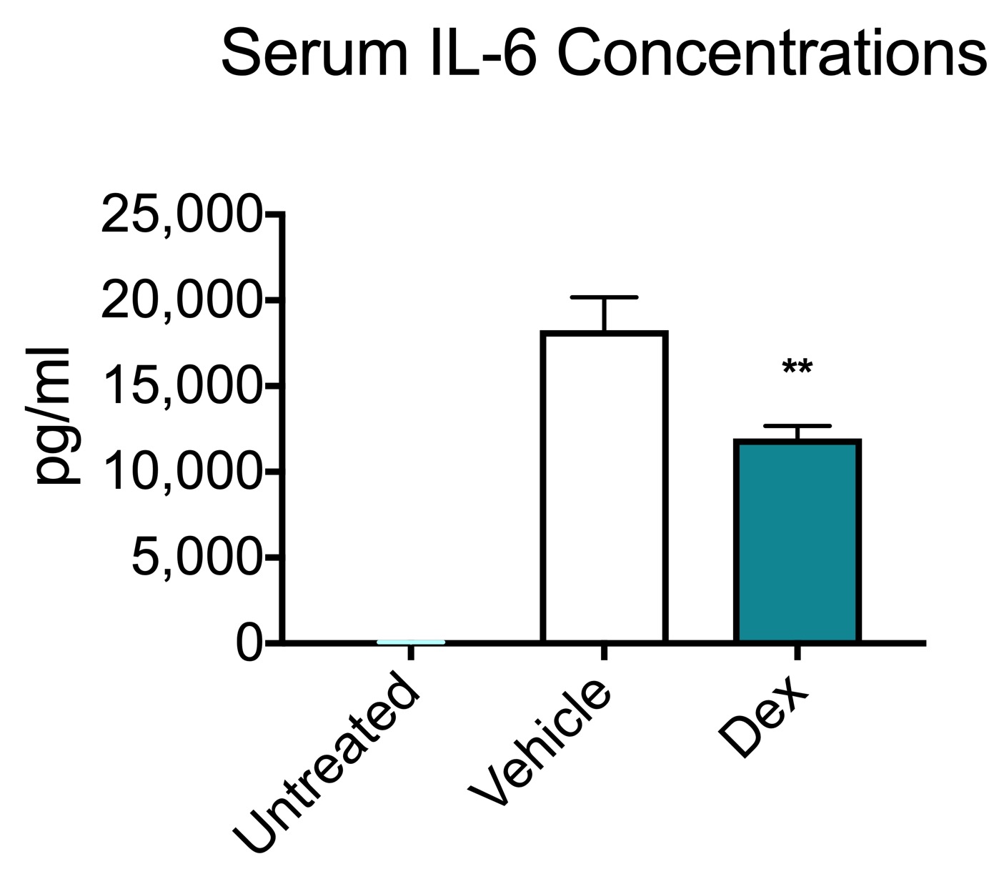
Suppression of LPS-induced inflammatory responses by the hydroxyl groups of  dexamethasone | Oncotarget
Suppression of LPS-induced inflammatory responses by the hydroxyl groups of dexamethasone | Oncotarget
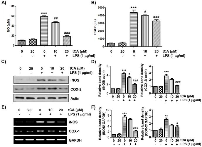
Inhibition of Lipopolysaccharide-Induced Inflammatory and Oxidative  Responses by Trans-cinnamaldehyde in C2C12 Myoblasts
Inhibition of Lipopolysaccharide-Induced Inflammatory and Oxidative Responses by Trans-cinnamaldehyde in C2C12 Myoblasts

Human lipopolysaccharide models provide mechanistic and therapeutic  insights into systemic and pulmonary inflammation | European Respiratory  Society
Human lipopolysaccharide models provide mechanistic and therapeutic insights into systemic and pulmonary inflammation | European Respiratory Society
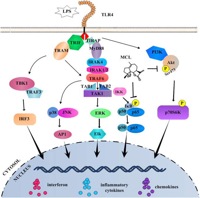
Micheliolide inhibits LPS-induced inflammatory response and protects mice  from LPS challenge | Scientific Reports
Micheliolide inhibits LPS-induced inflammatory response and protects mice from LPS challenge | Scientific Reports

RS-5645 attenuates inflammatory cytokine storm induced by SARS-CoV-2 spike  protein and LPS by modulating pulmonary microbiota
RS-5645 attenuates inflammatory cytokine storm induced by SARS-CoV-2 spike protein and LPS by modulating pulmonary microbiota
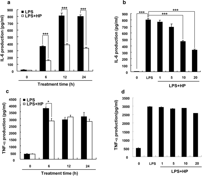
Anti-inflammatory effect of hispidin on LPS induced macrophage inflammation  through MAPK and JAK1/STAT3 signaling pathways | Applied Biological  Chemistry | Full Text
Anti-inflammatory effect of hispidin on LPS induced macrophage inflammation through MAPK and JAK1/STAT3 signaling pathways | Applied Biological Chemistry | Full Text

Single-cell secretion analysis reveals a dual role for IL-10 in restraining  and resolving the TLR4-induced inflammatory response | bioRxiv
Single-cell secretion analysis reveals a dual role for IL-10 in restraining and resolving the TLR4-induced inflammatory response | bioRxiv
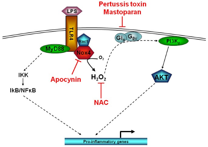
LPS induced inflammatory responses in human peripheral blood mononuclear  cells is mediated through NOX4 and Giα dependent PI-3kinase signalling |  Journal of Inflammation | Full Text
LPS induced inflammatory responses in human peripheral blood mononuclear cells is mediated through NOX4 and Giα dependent PI-3kinase signalling | Journal of Inflammation | Full Text

Alveolar macrophage phagocytic activity is enhanced with LPS priming, and  combined stimulation of LPS and lipoteichoic acid synergistically induce  pro-inflammatory cytokines in pigs - Mohammad Ariful Islam, Maren Pröll,  Michael Hölker, Ernst
Alveolar macrophage phagocytic activity is enhanced with LPS priming, and combined stimulation of LPS and lipoteichoic acid synergistically induce pro-inflammatory cytokines in pigs - Mohammad Ariful Islam, Maren Pröll, Michael Hölker, Ernst
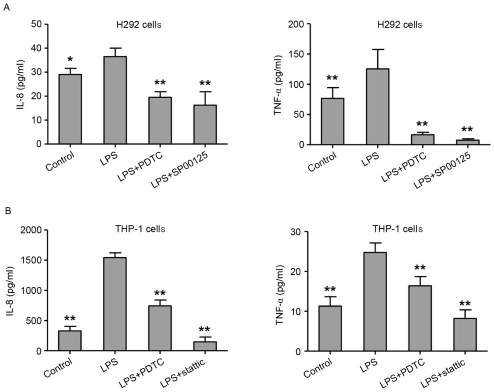
LPS‑induced proinflammatory cytokine expression in human airway epithelial  cells and macrophages via NF‑κB, STAT3 or AP‑1 activation
LPS‑induced proinflammatory cytokine expression in human airway epithelial cells and macrophages via NF‑κB, STAT3 or AP‑1 activation

Bavachin attenuates LPS-induced inflammatory response and inhibits the  activation of NLRP3 inflammasome in macrophages - ScienceDirect
Bavachin attenuates LPS-induced inflammatory response and inhibits the activation of NLRP3 inflammasome in macrophages - ScienceDirect

LPS Model of Systemic Inflammation - Melior Discovery

Noncanonical STAT1 phosphorylation expands its transcriptional activity  into promoting LPS-induced IL-6 and IL-12p40 production | Science Signaling
Noncanonical STAT1 phosphorylation expands its transcriptional activity into promoting LPS-induced IL-6 and IL-12p40 production | Science Signaling

Lipopolysaccharide-driven Th2 Cytokine Production in Macrophages Is  Regulated by Both MyD88 and TRAM*
Lipopolysaccharide-driven Th2 Cytokine Production in Macrophages Is Regulated by Both MyD88 and TRAM*

Skin culture induced acne model (LPS)

LPS-Induced Rodent Sepsis Model - Creative Biolabs

Inhibition of Lipopolysaccharide-Induced Inflammatory and Oxidative  Responses by Trans-cinnamaldehyde in C2C12 Myoblasts
Inhibition of Lipopolysaccharide-Induced Inflammatory and Oxidative Responses by Trans-cinnamaldehyde in C2C12 Myoblasts

Methane limit LPS-induced NF-κB/MAPKs signal in macrophages and suppress  immune response in mice by enhancing PI3K/AKT/GSK-3β-mediated IL-10  expression | Scientific Reports
Methane limit LPS-induced NF-κB/MAPKs signal in macrophages and suppress immune response in mice by enhancing PI3K/AKT/GSK-3β-mediated IL-10 expression | Scientific Reports
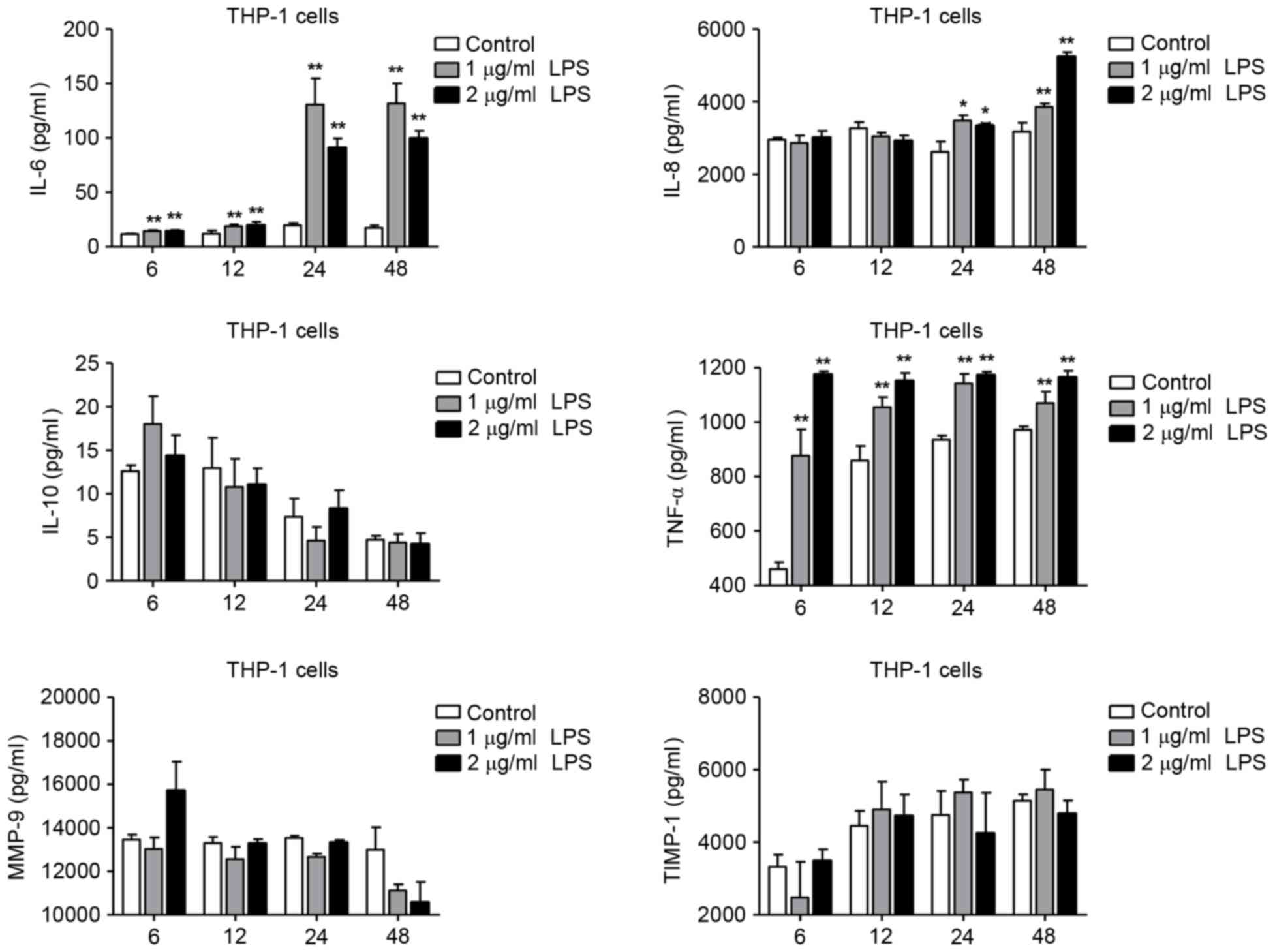
LPS‑induced proinflammatory cytokine expression in human airway epithelial  cells and macrophages via NF‑κB, STAT3 or AP‑1 activation
LPS‑induced proinflammatory cytokine expression in human airway epithelial cells and macrophages via NF‑κB, STAT3 or AP‑1 activation

Imoxin attenuates LPS‐induced inflammation and MuRF1 expression in mouse  skeletal muscle - Valentine - 2018 - Physiological Reports - Wiley Online  Library
Imoxin attenuates LPS‐induced inflammation and MuRF1 expression in mouse skeletal muscle - Valentine - 2018 - Physiological Reports - Wiley Online Library

LPS-induced TNF-α factor (LITAF)-deficient mice express reduced LPS-induced  cytokine: Evidence for LITAF-dependent LPS signaling pathways | PNAS
LPS-induced TNF-α factor (LITAF)-deficient mice express reduced LPS-induced cytokine: Evidence for LITAF-dependent LPS signaling pathways | PNAS
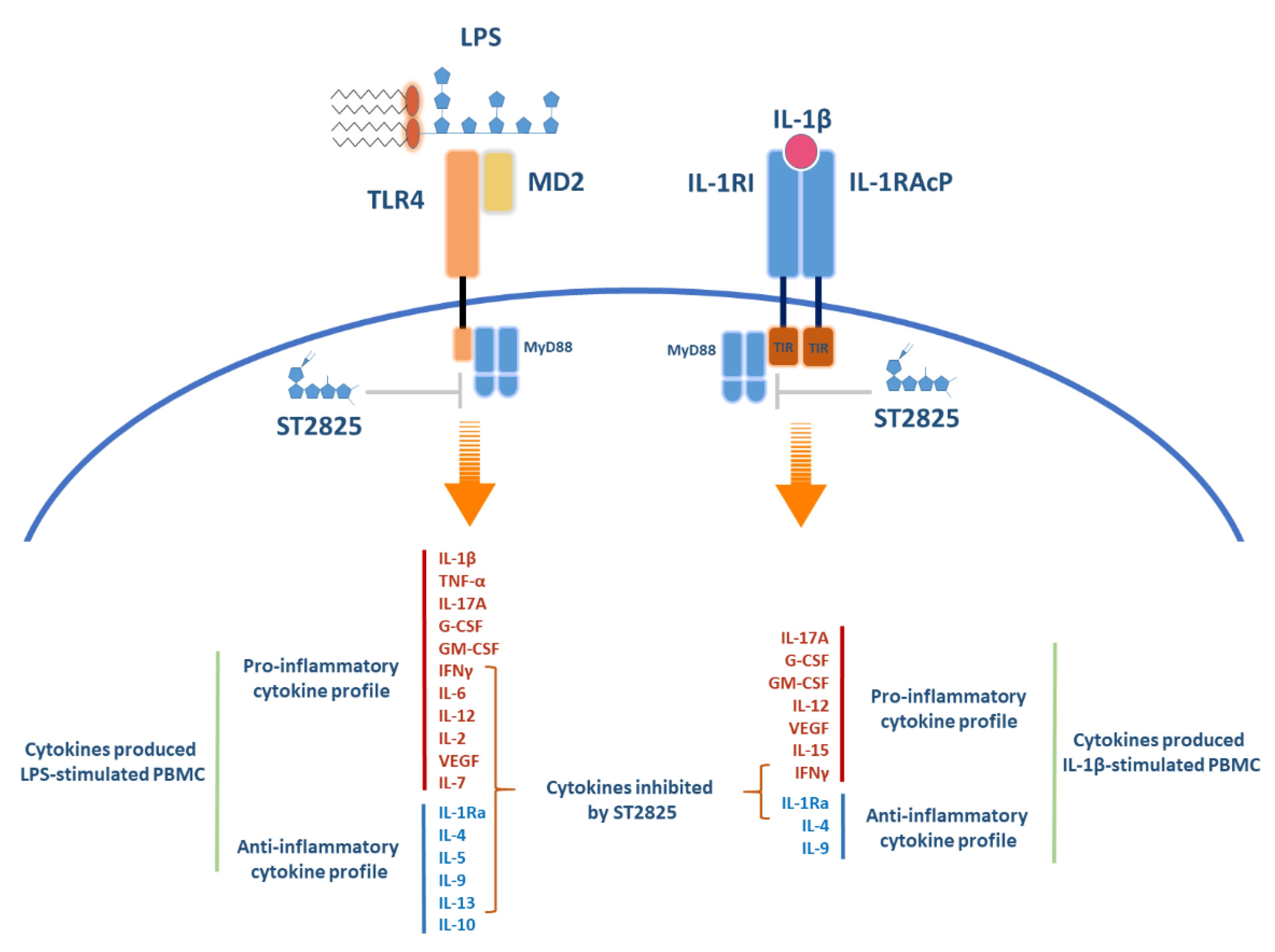
Molecules | Free Full-Text | Downregulation of Inflammatory Cytokine  Release from IL-1β and LPS-Stimulated PBMC Orchestrated by ST2825, a MyD88  Dimerisation Inhibitor
Molecules | Free Full-Text | Downregulation of Inflammatory Cytokine Release from IL-1β and LPS-Stimulated PBMC Orchestrated by ST2825, a MyD88 Dimerisation Inhibitor
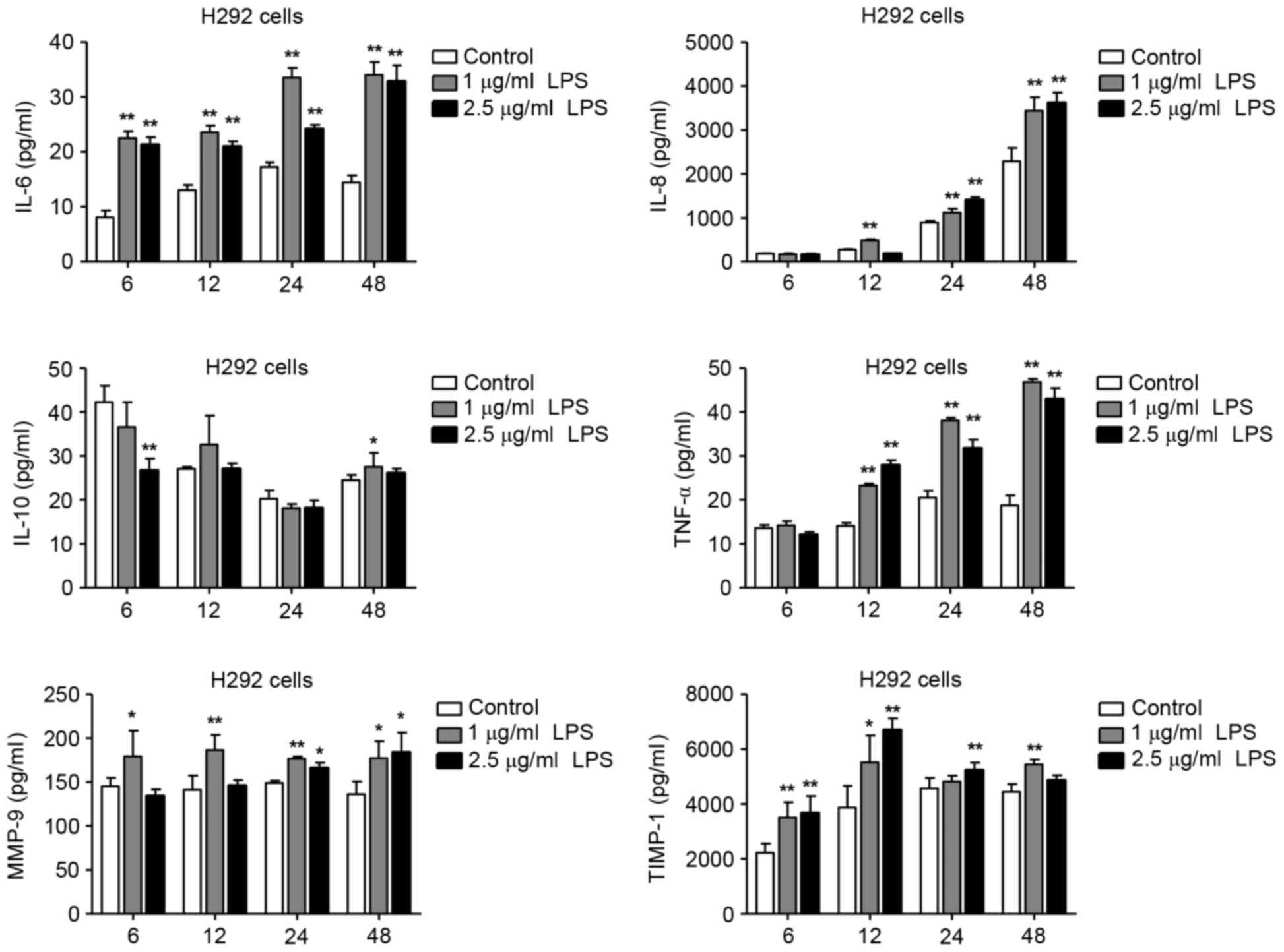
LPS‑induced proinflammatory cytokine expression in human airway epithelial  cells and macrophages via NF‑κB, STAT3 or AP‑1 activation
LPS‑induced proinflammatory cytokine expression in human airway epithelial cells and macrophages via NF‑κB, STAT3 or AP‑1 activation